A multi-dimensional detection system is employed to confirm the characteristics of
exosomes: transmission electron microscopy (TEM) is used to observe their characteristicstructures, nanoparticle analyzers or nano-flow cytometers are utilized to detect particle sizedistribution, and Western blot is performed to verify marker proteins such as CD9/CD63. Simultaneously, sequencing technology is applied to analyze nucleic acid composition, andmass spectrometry is used to identify protein components, ensuring that exosome samples
meet international research standards and providing a solid data foundation for subsequent
experiments.
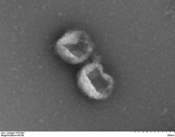
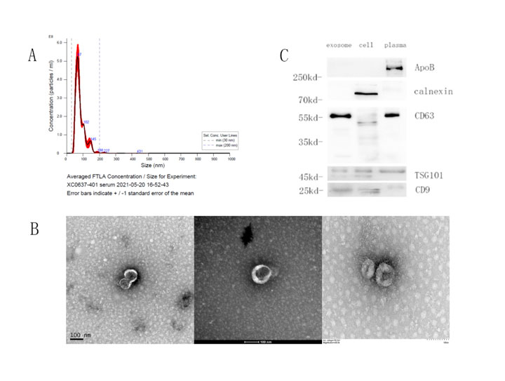
Electron microscopy images show
Display of exosome identification results
(Figure A shows the NTA particle size distribution, Figure B presents the WB protein marker
identification results, and Figure C depicts the morphological structure of exosomes
captured by electron microscopy.)
0512-69885880 商务合作:13911441296/18625299566
wutl@carrierbiomed.com
AI Industrial Park, No. 88 Jinjihu Avenue,Suzhou Industrial ParkE2-201